Advanced Upper Cervical Imaging Evaluation
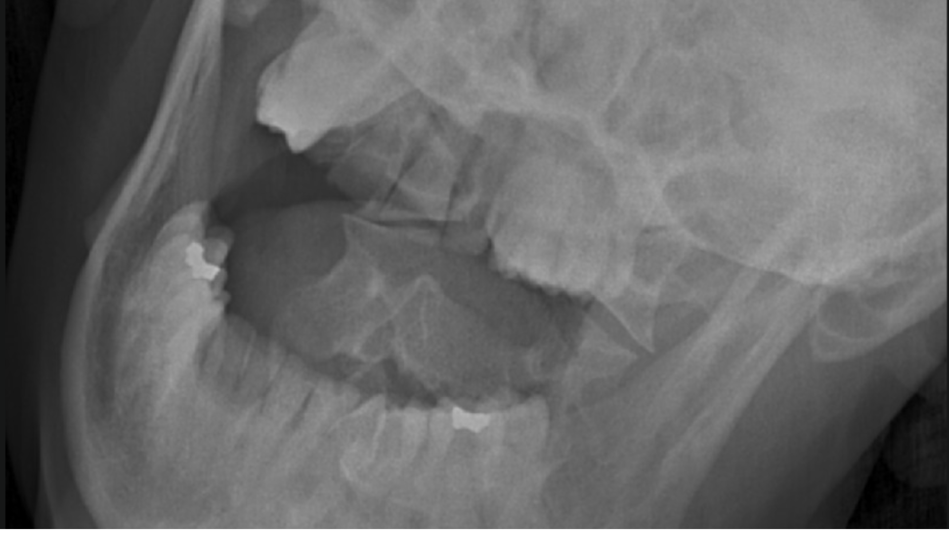
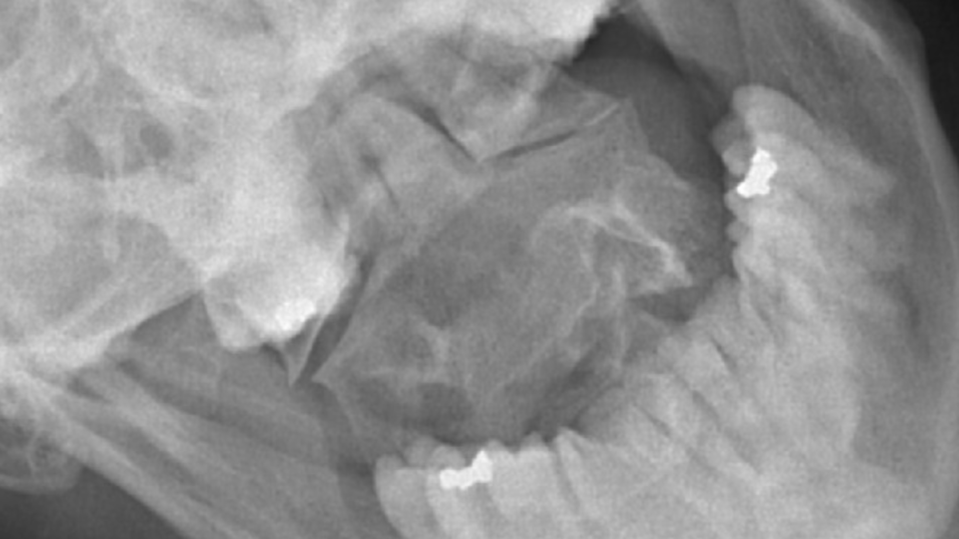
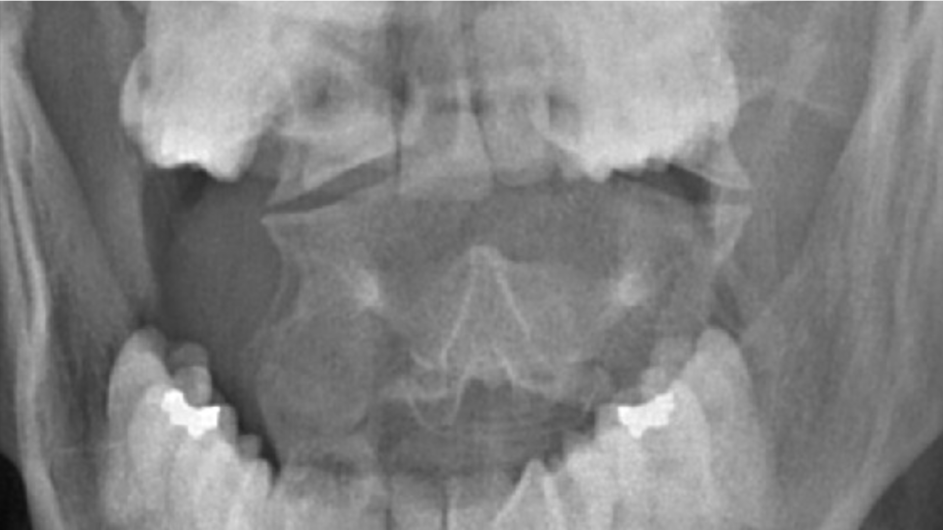
Davydov Pain Center provides advanced craniocervical junction (CCJ) imaging evaluation focused on assessment of increased upper cervical motion, cervical alignment, and upper cervical biomechanics.
Diagnostic imaging may assist evaluation of cervicocranial instability (CCI) as a contributor to chronic headaches, dizziness, upper quarter pain, fatigue, neck discomfort, and posture-related symptoms.
Weight-bearing AP OM neck lateral flexion X-ray imaging evaluation has been described in medical literature as a gold standard diagnostic approach for assessment of CCI in the sagittal plane.
Upper cervical conditions may contribute to chronic headaches, dizziness, posture-related symptoms, neck discomfort, and TMJ dysfunction.
Evidence-Based Imaging Evaluation
In September 2024, Smartray® GLX 0.1 imaging data was admitted into evidence in a personal injury case before the Bronx Supreme Court.
The imaging findings contributed to an $800,000 settlement in a case that was about to be lost by plaintiff.
This represented the first known court admission of Smartray® cervicocranial imaging data as diagnostic medical evidence, highlighting the importance of reproducible and reliable imaging evaluation.
“Weight bearing gold standard CCI diagnosis.”
Bronx Supreme Court Index #29770/2017e
The Gold Standard for CCI in Sagittal Plane
“AP OM neck lateral flexion weight bearing X-ray imaging study is considered a gold standard diagnostic test for determining CCI in the sagittal plane.”
“Occult Hypermobility of the Craniocervical Junction: A Case Report and Review.” Journal of Orthopaedic & Sports Physical Therapy. June 2011 | Volume 41 | Number 6 | Page 444
The Cervicocranium is One of the Most Difficult Anatomies to Evaluate Radiographically
The cervicocranium extends from the skull base through the second cervical intervertebral disk and represents one of the most complex anatomical regions in diagnostic imaging evaluation.
“Of the entire cervical spine, the Cervicocranium is the most difficult to evaluate radiographically because of its complex anatomy, physiologic motions, and normal variants that may simulate acute injury.”
Medical literature also describes the inherent difficulty in obtaining diagnostic frontal and lateral imaging projections because of subtle radiographic findings and complex upper cervical biomechanics.
“Cervicocranium: Its Radiographic Assessment.” John H. Harris Jr, MD, DSc. Radiology 2001; 218:337-351.
Request Your Consultation
Contact Davydov Pain Center to request an appointment for advanced CCI and TMJ imaging evaluation and upper quarter pain assessment.
Schedule Appointment